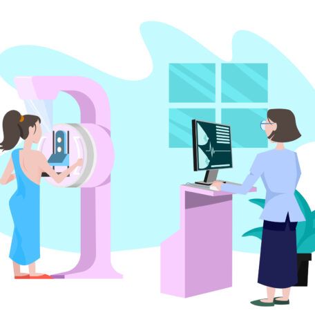
Mammograms save lives. They are the best way to detect breast cancer early, sometimes up to three years before abnormalities can be felt.
Our Approach to Mammograms
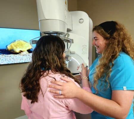
To ensure you feel comfortable during your mammogram, we use new technology like the Pristina Dueta™ patient-assisted compression device, giving patients control and comfort in their screening experience without sacrificing the clarity and quality of the image.
Our centers also offer Sensory Suite, which uses imagery, scent, and sound to enhance the mammogram experience.
3D Mammography
Our 15 Kettering Health Breast Centers offer 3D Mammography. A 3D mammogram takes multiple images of the breast to create a 3D image for the radiologist to see the breast tissue more clearly.
These take images of your breasts from many different angles. This allows the radiologist to look at breast tissue one layer at a time, making it easier to find abnormalities. This is especially helpful for women with dense breast tissue.
Additional Imaging
Following a mammogram, you may be called for a follow-up. All this means is that your radiologist wants to take a closer look with a more particular imaging procedure, such as additional mammography views, a breast ultrasound or, in some cases, a breast MRI.
An MRI usually comes last, and we would do additional mammography views or a breast ultrasound. If an abnormality is found, a biopsy might be recommended.
When should you receive a mammogram?
We follow the guidelines of the American College of Radiology, which states a woman should start having a mammogram at 40 years old and have them annually.
For those with a family history of breast cancer, they need to start having mammograms at an earlier age. It’s best to speak with a personal physician to find out when is right for you.
Types of mammograms
Screening mammograms are X-rays of the breasts done on a routine basis for women who do not exhibit any signs or symptoms of breast cancer.
Diagnostic mammograms are used to diagnose unusual breast changes, such as a lump, pain, nipple discharge, or change in breast size or shape. If your screening mammogram does show an abnormality, you may need additional imaging like a diagnostic mammogram. The radiologist will evaluate the images prior to a patient leaving the center so they understand the next steps.
New Law Expands Insurance for 3D Mammography, Dense Breasts
Read More


