Prostate cancer is one of the most common cancers in men.
Effectively treating prostate cancer, whether a new diagnosis or if it’s come back (known as “recurrent”), requires knowing where the cancer is located. With precise testing, your physician can prescribe the best treatment for getting you back to your best health.
PSMA imaging helps us do that.
What is PSMA-targeted PET?
Prostate Specific Membrane Antigen (PSMA) tracers are FDA-approved radioactive drugs used with PET and CT imaging. The PSMA bind to prostate cancer cells and “light up” the cancer cells on a scan to show doctors precisely where prostate cancer is located.
PSMA-targeted PET helps detect prostate cancer early and can be part of a complete cancer diagnosis by your physician.
Do I need a PSMA-targeted PET scan?
The FDA currently approves this type of imaging for
- Patients who have high-risk disease and are candidates for initial definitive therapy.
- Patients with suspected recurrence based on elevated levels of prostate-specific antigen (PSA) level.
The National Comprehensive Cancer Network recommends PSMA-targeted PET scans as a tool to help diagnose and manage prostate cancer.
How does it work?
Recognized as a “target” to help diagnose and treat prostate cancer cells decades ago, PSMA is a great “biomarker” for prostate cancer. They are expressed on normal prostate tissue cells. On prostate cancer cells, they’re overexpressed. This makes them helpful indicators for prostate cancer.
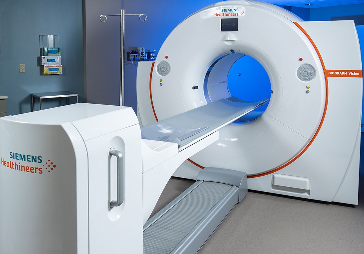
These scans work by attaching a radioactive tracer (called a positron-emitting isotope) to the PSMA. The PET scan machine detects the tracer and creates 3D images of a patient’s body, highlighting the prostate cancer cells.

Recent developments have refined imaging techniques to give us tracers such as F-18 and Ga-68 PSMA. These tracers in PET scan imaging guide diagnosis and treatment decisions by physicians caring for patients with prostate cancer.
The next step in development is adding a radioisotope that would allow the treatment of advanced prostate cancer by using the same targeting methodology as imaging, but now delivering therapy specifically targeting cancerous prostate cells.
PSMA PET Imaging explained by trusted expert Robert Flavell, MD, PhD
What is the difference?
PSMA PET/CT is more accurate than traditional imaging, such as Nuclear Medicine Bone scan and CT scan.
Contrasted with conventional imaging (CT, MRI), a this scan detects disease at initial staging and recurring metastatic disease with more sensitivity and specificity.
What happens during the scan?
The day before your scheduled PSMA PET/CT, a nurse will call you to ask you about your medications and remind you of your appointment time. The exam doesn’t require fasting. We encourage patients to drink fluids before and after their exam.
After arriving for your scan, we will ask for your height and weight. You’ll also need to remove metal objects such as jewelry and belt buckles. A nurse will place an IV in your arm. And a technologist will use the IV to inject radioactive PSMA. We will remove the IV after the injection. You’ll wait in a comfortable room for an hour while the PSMA circulates and attaches to prostate cancer cells.


