Cardiac PET at Kettering Health
Kettering Health offers comprehensive imaging services to diagnose heart disease, including a cardiac PET stress test.
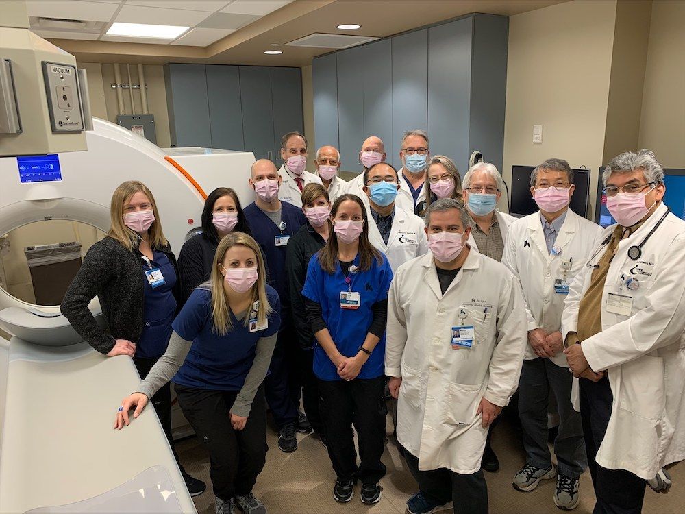
At Kettering Health Main Campus, we provide the only PET imaging service in western Ohio. With more than 30 years of experience in research and use of cardiac PET imaging, we proudly offer this non-invasive imaging test so you can find the care you need close to home.
What Is a PET Stress Test?
Positron emission tomography (PET) is the newest and most accurate non-invasive tool your doctor has to diagnose heart disease.
Your doctor may order what’s called a PET myocardial perfusion imaging scan, commonly known as a PET stress test or a Rubidium PET.
This test shows how well blood flows to your heart’s muscles while you’re at rest and during stress. Your doctor will compare the two images to see if there are any blockages.
What Is the Difference Between a Nuclear Medicine Stress Test and a PET Stress Test?
Since the 1970s, nuclear medicine stress tests have been used to image the heart, including at Kettering Health’s medical facilities.
These exams use SPECT (single photon emission computed tomography) to create a picture of your heart, using a small amount of radiation. But these exams have limits in their accuracy, especially for patients who carry more weight or have dense breast tissue.
Getting a PET stress test means a shorter exam and lower radiation exposure for patients. And PET stress tests offer what’s called coronary flow reserve calculations. These measure how much blood flows to the muscles of the heart.
PET testing uses positron tracers and a PET camera to create a 3D image of your heart. The tracers help create a clear image, unaffected by a patient’s weight or breast-tissue density.
What Happens during the PET Stress Test?
Before your exam, you will be asked not to eat or drink anything for 4 hours before your appointment. You may have small drinks of water. Other instructions include:
- Not eating or drinking anything containing caffeine (chocolate, soda, energy drinks, tea, coffee, Excedrin) 24 hours before your exam.
- Bringing a list of all your medications and speaking with your physician about stopping medications before your test.
- Wearing loose, comfortable clothing the day of your test.
- Your doctor may ask you to hold certain medications including beta blockers, nitrates, dipyridamole, theophylline or any medications containing caffeine.
When you arrive, a nurse or technologist will ask questions about your medical history and explain the test. He or she will also start a small IV in your arm.
The test will be performed on a flat imaging table with your arms above your head. The exam will last for 30-45 minutes, and a physician will monitor the exam. If feel you cannot keep your arms above your head, please let your nurse or technologist know.
After the exam, a specialized imaging doctor will review the pictures. The doctor will then send the results to your ordering physician and make them available in MyChart.


